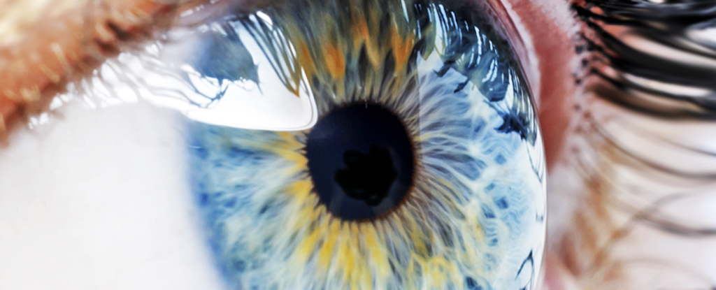
In efforts to tackle the leading cause of blindness in developed countries, researchers have recruited nanotechnology to help regrow retinal cells.
Macular degeneration is a form of central vision loss, which has massive social, mobility, and mental consequences. It impacts hundreds of millions of people globally and is increasing in prevalence.
The degeneration is the consequence of damaged retinal pigment cells. Our bodies are unable to grow and replace these cells once they start dying, so scientists have been exploring alternative methods to replace them and the membrane within which they sit.
“In the past, scientists would grow cells on a flat surface, which is not biologically relevant,” explains Anglia Ruskin University biochemist Barbara Pierscionek.
“Using these new techniques the cell line has been shown to thrive in the 3D environment provided by the scaffolds.”
Nottingham Trent University biomedical scientist Biola Egbowon and colleagues fabricated these 3D scaffolds with polymer nanofibers and coated them with a steroid to reduce inflammation.
Using a technique called electrospinning, which produces nanometer-wide fibers by squirting a molten polymer through a high-voltage field, the team was able to keep the scaffold sufficiently thin.
The polyacrylonitrile polymer they used provided mechanical strength, and Jeffamine polymer attracts water, essentially allowing the synthetic scaffold to act as a membrane.
The water-attracting ability of the material is what helps the cells bind to the scaffold and also encourages their growth, but when the effect is too strong, it’s also been associated with cell death in previous research.
The team’s new formulation seems to be just right, as the system increased the growth and longevity of the retinal lab cells and kept them viable for at least 150 days.
“This research has demonstrated, for the first time, that nanofiber scaffolds treated with the anti-inflammatory substance such as fluocinolone acetonide can enhance the growth, differentiation, and functionality of retinal pigment epithelial cells,” says Pierscionek.
Previous attempts have used collagen and cellulose to create a similar scaffold, but Egbowon and team believes their synthetic option will be easier to make compatible with our immune systems and simpler to modify.
The new study has demonstrated this method can keep the required single layer of retinal cells healthy, producing biomarkers that indicate they are functioning more naturally than what has been found when they grow on other mediums.
However, there’s still a lot we don’t know about how viable this approach will be for treating human patients with macular degeneration.
“While this may indicate the potential of such cellularized scaffolds in regenerative medicine, it does not address the question of biocompatibility with human tissue,” Egbowon and colleagues caution in their paper, as there is a massive difference between growing cells in a petri dish and having a functioning tissue substitute within a body.
Other research in this area is already investigating whether lab grown cells can be plugged back into other retinal cell types to form functioning units of tissue. Another tactic involves activating cells already in human eye tissues that regenerate retinal cells in other animals.
The team’s next steps will be to investigate the orientation of the cells, which is important for ensuring they can maintain a good blood supply, before they can be considered for testing inside a living system.
This research was published in Materials & Design.


